Shared posts

14 Nov 10:38

Simulation and Understanding

by Derek Lowe

Roald Hoffmann and Jean-Paul Malrieu have a three-part essay out in Angewandte Chemie on artificial intelligence and machine learning in chemistry research, and I have to say, I’m enjoying it more than I thought I would. I cast no aspersion against the authors (!) – it’s just that long thinkpieces from eminent scientists, especially on such broad topics as are covered here, do not have a good track record for readability and relevance. But this one’s definitely worth the read. It helps that it’s written well; these subjects would be deadly – are deadly, have been deadly – if discussed in a less immediate and engaging style. And if I had to pick a single central theme, it would be the quotation from René Thom that the authors come back to: prédire n’est pas expliquer, “To predict is not to explain”.

The first part is an introduction to the topics at hand: what do we mean when we say that we’ve “explained” something in science? The authors are forthright:

To put it simply, we value theory. And simulation, at least the caricature of simulation we describe, gives us problems. So we put our prejudices up front.

They place theory, numerical simulation, and understanding at the vertices of a triangle. You may wonder where experimental data are in that setup, but as the paper (correctly) notes, the data themselves are mute. This essay is about what we do with the experimental results, what we make of them (the authors return to experiment in more detail in the third section). One of their concerns is that the current wave of AI hype can along the way demote theory (a true endeavor of human reason if ever there was one) to “that biased stuff that people fumbled along with before they had cool software”.

Understanding is a state of mind, and one good test for it is whether you have a particular concept or subject in mind well enough, thoroughly enough, that you can bring another person up to the same level you have reached. Can you teach it? Explain it so that it makes sense to someone else? You have to learn it and understand it inside your own head to do that successfully – I can speak from years of experience on this very blog, because I’ve taught myself a number of things in order to be able to write about them.

My own example of what understanding is like goes back to the Euclid’s demonstration that the number of primes is infinite (which was chosen for this purpose by G. H. Hardy in A Mathematician’s Apology). A prime number, of course, is not divisible by any smaller numbers (except the universal factor of 1). So on the flip side, every number that isn’t prime is divisible by at least one prime number (and usually several) – primes are the irreducible building blocks of factors. Do they run out eventually? Is there a largest prime? Euclid says, imagine that there is. Let’s call that number P – it’s a prime, which means that it’s not divisible by any smaller numbers, and we are going to say that it’s the largest one there is.

Now, try this. Let’s take all the primes up to P (the largest, right?) and multiply them together to make a new (and rather large) number, Q. Q is then 2 · 3 · 5 · 7 · 11 · (lots of primes all the way up to) · P. That means that Q is, in fact, divisible by all the primes there are, since P is the last one. But what happens when you have the very slightly larger number Q+1? Now, well. . .that number isn’t divisible by any of the primes, because it’ll leave 1 as a remainder every single time. But that means that Q+1 is either divisible by some prime larger than P, or it’s a new prime itself (and way larger than P) and we just started by saying that there aren’t any such things. The assumption that P is the largest prime has just blown up; there are no other options. There is no largest prime, and there cannot be.

As Hardy says, “two thousand years have not written a wrinkle” on this proof. It is a fundamental result in number theory, and once you work your way through that not-too-complicated chain of reasoning, you can see it, feel it, understand that prime numbers can never run out. The dream is to understand everything that way, but only mathematicians can approach their subject at anything close to that level. Gauss famously said that if you didn’t immediately see why Euler’s identity (e +1 = 0) had to be true, then you were never going to be a first-rate mathematician. And that’s just the sort of thing he would say, but then again, he indisputably was one and should know.

Now you see where Thom is coming from. And where Hoffmann and Malrieu are coming from, too, because their point is that simulation (broadly defined as the whole world of numerical approximation, machine-learning, modeling, etc.) is not understanding (part two of their essay is on this, and more). It can, perhaps, lead to understanding: if a model uncovers some previously unrealized relationship in its pile of data, we humans can step in and ask if there is something at work here, some new principle to figure out. But no one would say that the software “understands” any such thing. People who are into these topics will immediately make their own prediction, that Searle’s Chinese Room problem will make an appearance in the three essays, and it certainly does.

This is a fine time to mention a recent result in the machine-learning field – a neural-network setup that is forced to try to distill its conclusions down to concise forms and equations. The group at the ETH working on this fed it a big pile of astronomical observations about the positions of the planet Mars in the sky. If you’re not into astronomy and its history like I am, I’ll just say that this puzzled the crap out of people for thousands of years, because Mars does this funny looping sewing-stitch-like motion in the sky, occasionally pausing among the stars and then moving backwards before stopping yet again and resuming its general forward motion across the sky). You can see why people starting added epicycles to make this all come out right if you started by putting the Earth at the center of the picture. This neural network, though, digested all this and came up with equations that, like Copernicus (whose birthday I share), puts the sun at the center and has both Earth and Mars going around it, with us in the inner orbit. But note:

Renner stresses that although the algorithm derived the formulae, a human eye is needed to interpret the equations and understand how they relate to the movement of planets around the Sun.

Exactly. There’s that word “understand” again. This is a very nice result, but nothing was understood until a human looked at the output. When we start to wonder about that will be the day we can really talk about artificial intelligence. And to Hoffman and Marlieu’s point about simulation, it has to be noted that some of those epicycle models did a pretty solid job of predicting the motion of Mars in the sky. Simulations and models can indeed get the right numbers for the wrong reasons, with no way of ever knowing that those reasons were wrong in the first place, or what a “reason” even is, or what is meant by “wrong”. And the humans that came up with the epicycles knew that prediction was not explanation, either – they had no idea why such epicycles should exist (other than perhaps “God wanted it that way”). They just knew that these things made the numbers come out right and match the observations, the early equivalent of David Mermin’s quantum-mechanics advice to “shut up and calculate“.

Well, “God did it that way” is the philosophical equivalent of dividing by 1: works every time, doesn’t tell you a thing. We as scientists are looking for the factors in between, the patterns they make, and trying to work out what those patterns tell us about still larger questions. That’s literally true about the distribution of prime numbers, and it’s true for everything else we’ve built on top of such knowledge. In part three of this series of essays, the authors say

The wave of AI represented by machine learning and artificial neural network techniques has broken over us. Let’s stop fighting, and start swimming. . .We will see in detail that most everything we do anyway comes from an intertwining of the computational, several kinds of simulation, and the building of theories. . .

They end on a note of consilience. As calculation inevitably gets better, human theoreticians will move up a meta-level, and human experimentalists will search for results that break the existing predictions. It will be a different world than the one we’ve been living in, which frankly (for all its technology) will perhaps come to look more like the world of Newton and Hooke than we realize, just as word processing and drawing programs made everything before them seem far more like scratching mud tablets with sticks and leaving them to dry in the sun. We’re not there yet, and in many fields we won’t be there for some time to come. But we should think about where we’re going and what we’ll do when we arrive.

04 Nov 13:29

Hindered Ethers Made Easier

by Derek Lowe

Since I mentioned a new Mitsunobu-type reaction yesterday, I should note that a new route to hindered ethers has come out this summer from the Baran group at Scripps. Here’s the ChemRxiv version, and here’s the Nature paper that just appeared. And there are more details at the group’s blog here. It’s an electrochemical reaction that involves decarboxylation to give very reactive carbocations that are then trapped by oxygen nucleophiles. You can make ethers, or use the reaction as a way to prepare alcohols if you just let water trap the cation, and if you really are into it, you can even trap with things like fluoride (albeit in lower yields).

These kinds of hindered products are totally unavailable through Mitsunobu chemistry, or through the classic Williamson ether synthesis, or basically anything that involves Sn2 nucleophilic attack. And the paper shows some dramatic examples of this, with compounds that were either unknown or only prepared by all-the-way-around-the-barn methods that can now be made directly. If you read the paper, you’ll see that this took a lot of experimentation. There are numerous variables and side reactions, and you cannot figure out how to solve them from first principles. The Kolbe electrolysis reaction is the 19th-century forerunner (1847!) to this work and the related Hofer-Moest reaction (1902) is an even more direct ancestor. But that uses the alcohol partner as the solvent, which is not a practical solution:

It became immediately apparent that limiting the amount of alcohol posed several considerable challenges: the decomposition of the carbocation due to the low nucleophilicity of alcohols; the competitive trapping of the carbocation by water; the consumption of alcohols by anodic oxidation; and the necessity of an external electron-acceptor in order to balance electrons. Figure 1c summarizes the results of around 1,000 experiments (see Supplementary Information for an extensive sampling) that were undertaken in order to solve these problems.

Adjusting the solvent, the electron acceptor, the anode material, and the addition of small amounts of base were all crucial, but the resulting reaction looks pretty robust (and naturally can be realized with the Baran-developed ElectraSyn device). Olefins, esters, acetals, Boc- and Cbz-amines, heterocycles, alkyl and aryl halides, and boronic esters are all compatible with the conditions. As the blog post linked above notes, though, you’ll do best if your acid (and the resulting carbocation) is tertiary, given the well-known stability order of carbocations.

So this could open up a number of compounds that normally you wouldn’t even think about, because ether synthesis is just synonymous in most synthetic chemists’ minds with nucleophilic displacement, and hindered things Just Don’t Work. We’ve all pushed the boundaries of such reactions. When I first started out in the lab in grad school, I had some tetrahydropyran derivatives to make, and a suggested synthetic route had been mapped out for me. I plunged in, but pretty quickly came up against the step to form the ring, which was an arrow with “Mitsunobu” written across the top. My first Mitsunobu! And it was hopeless; the starting material was just too hindered, as I shortly realized. I’ve told the story of how I got around that problem here, which was a valuable lesson in taking people’s word for stuff.

I would assume that intramolecular variants of this new electrochemical route would be a nice way to prepare crowded cyclic ethers as well. I’m always glad to see things come up that demolish the classic synthetic routes and disconnections that we all have in our heads!

04 Nov 13:07

A "living drug" that could change the way we treat cancer | Carl June

by contact@ted.com (TED)
Carl June is the pioneer behind CAR T-cell therapy: a groundbreaking cancer treatment that supercharges part of a patient's own immune system to attack and kill tumors. In a talk about a breakthrough, he shares how three decades of research culminated in a therapy that's eradicated cases of leukemia once thought to be incurable -- and explains how it could be used to fight other types of cancer.
30 Oct 15:30

How Close Do You Get to the Best Compound?

by Derek Lowe

Here’s a topic that came up in my Twitter feed the other day – I fear it’s unanswerable, but I’d like to hear what people have to say about it. Drug discovery projects start, of course, from a selection of possible chemical matter and chemical series, and they eventually narrow down to a clinical candidate. Various possibilities are given an airing along the way, but the one that makes it through has to come reasonably close to satisfying a whole list of criteria – potency, selectivity, metabolic stability, toxicology, ease of formulation, and others. Some of these can be rather closely coupled, while others are unrelated to each other, and others may be actually opposed (for example, the structural changes that bring on more potency might be just the ones that lead to worse pharmacokinetics).

One question is, though, how many other compounds that would be “development-worthy” are still in there when the project finishes up? That could include ones that were made near the end and didn’t get a full hearing, but I think the question is more directed at analogs that never got made at all. I would guess that most projects could in theory be squeezed for another clinical candidate from their same chemical series – many times this might come from something that is more difficult to make at the bench and was thus not followed.

This is related to a question that’s come up around here before: if you took the same new drug target and set several organizations to working on it at the same time, with their own chemical screens and their own set of medicinal chemists, how many different chemotypes would result? That experiment has been done, quite a few times, under natural conditions, by companies working on the same target but not knowing what the competition was up to. (Coming at a target when you know something about the competing chemical matter is a different case – you’re deliberately trying to avoid the competing patent claims, and you’ll have made some of the other stuff as a comparison for your assays). But in the flying-blind situation, it seems to depend on what sort of SAR the target protein will tolerate. There are cases (such as the PPARs) where the binding pocket accommodates all sorts of stuff and a wide variety of chemical matter shows up, and others where things are so constrained that some common features are almost inevitable. There may be a narrow tunnel in the binding site, or a metal atom that you pretty much have to coordinate to, or a basic region that any potent compound is going to have an acidic group to match with, and so on,

The specific question that came up on Twitter, though, is an even higher bar: how many existing drugs have even *better* variations that just didn’t get pursued? I suppose an alternate way of asking this would be, if you could completely set aside worries about patent claims, how many existing drugs could be re-worked by varying their existing structures to find something better? That immediately suggests the follow-up question of “What do you mean by better?”, and that’s going to be different for different drugs. Could be better selectivity, longer half-life, more (or perhaps less!) potency, avoiding some specific metabolite, etc. There are some drugs that you look at and say “Actually, that one doesn’t really have so many problems”, but there are plenty of others that could in theory be tweaked to something a bit better.

I think the closest thing we have to a real-world example of this is the first wave of deuterated drug analogs. The idea there was that compounds whose biggest problem was metabolism (short half-life or a metabolite best avoided) could be improved by selective deuteration to slow down specific enzymatic bond-breaking events. And at the time, no one wrote their patent language to accommodate such deuterated analogs, so IP-wise the field was pretty open. That story continues: Teva got the first approval in 2017 for a deuterated version of an older compound. Concert Pharmaceuticals is a major player in that area, and they just announced clinical results the other day for a deuterated version of Incyte’s ruxolitinib. So the idea works, but at the same time it hasn’t revolutionized the drug industry, either.

And that’s my guess about the answer to the “better variations” question. I think that clinical candidates (and especially marketed compounds) are certainly at the far end of the big distribution, and that most improvements would probably be small. If a major liability is uncovered, that almost always happens in time for a program to either be killed or to reset and search for newer, better chemical matter. So what makes it out the far end is already pretty good – I think it’s unlikely that there are too many notably better compounds (for the same target) to be made by variations on known drug structures. Thoughts?

30 Oct 13:54

Trifluoromethyl Amides, Now Available

by Derek Lowe

Early-stage medicinal chemists are going to be all over this paper that’s just come out in Nature. That’s because it opens up a whole interesting class of molecules that we’ve never really had access to: N-trifluoromethyl amides. That phrase won’t do much for you unless you’re a synthetic organic chemist, and especially one doing drug discovery work, but here’s why it’s a big deal.

Amides are everywhere, for starters. Amide bonds are what stitch amino acids into proteins, and the number of other biomolecules with amide functional groups is probably beyond counting, too. As you’d imagine, it’s a widely used motif in synthetic drug molecules as well, not least because forming garden-variety amides is one of the easiest and most reliable reactions known to science. If you have an amine group that you’d like to functionalize in this way, the number of carboxylic acids available to you is huge, and if you have a carboxylic acid, the same goes for the number of commercial amines. Amide formation is such an obvious way to crank out huge numbers of compounds that it’s long since become a cliché in the business, as in “I don’t want to just see five hundred amides coming out of this series”.

Going from a secondary amide (where there’s still an NH) to a tertiary one (where there are two carbons on the nitrogen) is a big switch. You can see that from the protein world; the only one of the canonical amino acids that has two substituents on its amine is proline. A simple methyl group on the nitrogen changes things – the polarity of the group, its hydrogen-bonding character, the rotation around the relevant bonds, and its stability against the (very wide) variety of enzymes that can break amides back down again. So N-methylation is one of the classic ways to modify a known peptide and turn it into something that might be unnatural enough to hang around longer in the gut or the bloodstream; it’s really one of the first things you do.

The medicinal chemists in the crowd know all this well, and they also know how important fluorinated compounds are in the business. I’ve gone on about them several times here, too, of course: fluorine is nearly the size of a plain hydrogen substituent, but is wildly different electronically. It pulls electron density out of whatever it’s attached to (which can change the character of things very much), its polarity gives it odd and useful interaction properties with all sorts of other functional groups, and the strength of the carbon-fluorine bond is legendary. If you see a C-H on your drug structure that’s being oxidatively metabolized and clearing out your drug candidate too quickly, the first thing you think of is whether that spot can be fluorinated, because the C-F analog will stop that in its tracks.

Now the big reveal: there has never been a good way to make N-trifluoromethyl amides. A few examples are known, but they’re been mostly one-offs. Those streams (N-methyl amides and fluorinated functional groups) have rarely crossed, because the N-CF3 group just ruins all those slick and easy ways to prepare amides. For starters, you can’t really get compounds containing the H-N-CF3 combination; they tend to be unstable, and if they can they’ll rip themselves apart through an elimination reaction and spit out HF, and nobody wants that. You wouldn’t expect that behavior so much from an N-trifluoromethyl amide, but it’s been hard to know, since they’re so hard to get to in the first place. Until now.

The paper linked above, from Franziska Schoenebeck and co-workers Thomas Scattolin and Samir Bouayad-Gervais at Aachen, provides an ingenious solution to the problem. As shown in the scheme, they start from an isothiocyanate (smelly, but widely available) and react that with silver fluoride. The group had already shown that this combination fluorinated the central carbon of such compounds, and in this case it goes all the way the a trifluoromethyl while stripping off the sulfur entirely. You’re left with that odd-looking trifluoromethylamine silver salt, which has the advantage of being stable enough to react cleanly with more silver fluoride and the phosgene equivalent bis(trichloromethyl) carbonate to give the acyl fluoride intermediate. Those, they found, are actually stable enough to store, which is good, because the final step gives you the diversity on the carbonyl end – reacting the acyl fluoride species with a Grignard (or probably several other sorts of organometallic reagents, I’d guess) gives you the desired N-trifluoromethyl amide, which you have been able to sneak up on without it realizing what you’re up to. Usefully, that final Grignard addition is fast enough that a bromoaryl group in your molecule will survive without doing metalation reactions of its own.

That acyl fluoride intermediate had also been known in a few examples in the literature, but the preps for such things tended to involve stoichiometric amounts of mercury salts and plenty of fluorophosgene gas, an extremely unappealing prospect. Silver fluoride and BTC is a lot easier system to deal with. Now, as the Nature “News and Views” piece on this paper notes, this whole procedure does go through five equivalents of silver fluoride on the way, which is not something that can be scaled up to drug-production levels. We early-stage research types can (and will!) use this system to explore this new world of functionality, but the world is going to need another way to make these things if we’re going to turn them into wonder drugs. (If someone has an idea for directly N-trifluoromethylating secondary amides, now’s the time to break it out. But that’s not going to be easy, since no one’s accomplished it already).

The paper shows a number of amino acid derivatives (chirality is retained, no problem) and many other structures containing aryls, heteroaryls, esters, sulfones, triflates, nitriles and so on. You can take the fluoroacyl intermediate and turn it into ureas and carbamates as well, which opens up still more possibilities. It turns out the the trifluoromethyl amides themselves are stable, can be taken through standard sorts of chemistry (such as Pd couplings) without reacting or falling apart, and show notably reduced barriers to bond rotation compared to the plain N-methyl analogs.

This work has application beyond medicinal chemistry, of course, materials science and polymer work being the first things that come to mind. But it’s for sure that no drug binding sites have ever laid eyes on a trifluoromethyl amide before. A lot of new compounds with unusual properties are going to get prepared pretty quickly, and it will be quite interesting to see what comes out!

29 Oct 13:55

[ASAP] Leveraging Atropisomerism to Obtain a Selective Inhibitor of RET Kinase with Secondary Activities toward EGFR Mutants

by Sean T. Toenjes†, Valeria Garcia†, Sean M. Maddox†§, Gregory A. Dawson†?, Maria A. Ortiz‡, F. Javier Piedrafita‡, and Jeffrey L. Gustafson*†

TOC Graphic

ACS Chemical Biology
DOI: 10.1021/acschembio.9b00407
29 Oct 13:03

Senescent cells feed on their neighbours

by Michael Overholtzer

Nature, Published online: 28 October 2019; doi:10.1038/d41586-019-03271-3

Chemotherapy-treated cancer cells that enter a non-dividing state called senescence can nevertheless boost cancer growth. The finding that these cells eat neighbouring cells reveals a mechanism that enables senescent cells to persist.
28 Oct 15:37

[ASAP] Nutrient-Based Chemical Library as a Source of Energy Metabolism Modulators

by Tomoyuki Furuta†, Yuya Mizukami‡, Lisa Asano†, Kenjiro Kotake†, Slava Ziegler§, Hiroki Yoshida†, Mizuki Watanabe†, Shin-ichi Sato†, Herbert Waldmann§, Makiya Nishikawa*‡?, and Motonari Uesugi*†?#

TOC Graphic

ACS Chemical Biology
DOI: 10.1021/acschembio.9b00444
22 Oct 15:11

Drugs Inside Cells: How Hard Can It Be, Right?

by Derek Lowe

One would imagine that we drug discovery and development types have a reasonable handle on how much of our latest candidate compound makes it into cells. And that we would further know how much of that is floating around freely in there, versus tied up to some protein or another. One could not be more wrong.

This is truly a major (the major?) unsolved problem in pharmacokinetics: intracellular concentrations and distribution. There are situations where we can get such data (often with a fluorescent compound or something of that sort), but those are far outnumbered by the ones where we really have only a fuzzy idea. Considering the number of drug targets that are inside the cell membrane, that’s a big gap – and we really should be saying “membranes” there, too, because once inside the cell you have the mitochondrial membrane, the nuclear membrane, and all sorts of other gates, walls, and velvet ropes that your compound might or might not pass.

This new paper (from Manchester) is a good look at the situation. As they say, understanding such things “would provide enormous clarity” which is now lacking. Unfortunately, the lack of clariy is not only due to the lack of data – we have our own misunderstandings to deal with, and the first one the paper calls out is the way that a lot of us think about compound binding (and free concentration) in the blood plasma:

A key driver of intracellular concentration in vivo must be the unbound drug present in the circulation. Unfortunately, this subject is greatly misunderstood in the scientific community. Prestige publications in high-impact international journals still carry a message that plasma protein binding determines the unbound concentration in the circulation. Anecdotal collection of publications and presentations would indicate that over one-half of the drug research scientific community has been misled.

Instead, it’s all about clearance – clearance of whatever got absorbed and escaped first-pass metabolism. Plasma protein binding is an equilibrium process, and the drain down at the bottom right of your mental picture of it is compound clearance. As the authors note, there are basically no examples of any drug-drug interaction that works through displacement of plasma protein binding. Doesn’t happen. If it really were a determining factor for unbound concentration, that wouldn’t be true.

There’s also confusion around the volume of distribution. Unconsciously (well, sometimes consciously) people assume that if a drug has a low distribution volume that it must also be reaching low concentrations inside cells, perhaps because it’s having difficulty reaching cells at all. But that doesn’t follow, either. The big influence on steady-state volume of distribution is compound binding (or, if you like, fraction of unbound compound, same thing). If your compound doesn’t bind to much, it’s going to show a high volume of distribution, since it’s going to be spread all over the aqueous compartment(s) of the body, and you can get all sorts of VoD numbers down from there as you get more or less lipophilic, charged, and so on, but these aren’t directly tied to unbound intracellular concentration.

The paper goes into several other pharmacokinetic assumptions of this sort; I definitely recommend it for clearing your head on these subjects. It then gets down to the question of intracellular concentrations, with examples from infectious disease (tuberculosis) and oncology, among others. Our data are just not all that good. As mentioned, many of the studies in the field depend on some sort of fluorescence, and tagging drug molecules with a fluorescent group profoundly changes their properties. You have cases like doxorubicin, where the drug itself has intrinsic fluorescence, but it’s pointed out that “dox” is likely a low-permeability drug to start with, and that the great majority of cellular permeability studies with it have been single-dose (with a relatively short time course) rather than multiple-dose steady-state conditions.

And getting such data without fluorescence is an even harder problem. The paper presents an analysis of published data on various tyrosine kinase inhibitors, and estimates that at best, most of them don’t have average steady-state concentrations even as high as four times the measured Ki or IC50 values, and if you assume a more physiological concentration of ATP, most of them probably have unbound plasma concentrations *below* those values. But they still work.

In the end, the paper presents two options to deal with all this uncertainty: we can try to come up with some new technology that can measure bound and unbound drug concentrations in different cell types (preferably in vivo). . .or we can just say the hell with it, assume that we need high (lipoidal) permeability to get through cell membranes, which will help to cancel out transporter effects in things like CNS, tumor cell, and bacterial penetration (and when possible, try to design away from those as well). The authors advocate the second choice! That’s largely because the technological barriers are extremely high. As they say, doing such measurements on single cells in homogeneous culture is still at or beyond the state of the art. And as soon as you move from single-cell measurements to samples that have different varieties of cells in them (that is, something approximating the real world), “the idea of technology helping to provide a global solution becomes almost fanciful“.

In the end, you’ll be trying to make compounds with the highest passive permeability you can, and avoiding the big efflux pumps like P-gP as much as possible. “But we already knew that”, is going to be the reaction, and I think that this paper is replying “Yes, you did, didn’t you? Or you should have.” The idea of measuring intracellular concentration (more particularly, unbound concentration, seems like something we would very much want, but “the more we ask the more complex it becomes“. Better, the authors say, to stick to first principles as much as you can.

I see their point! After all, those first principles are not going away, no matter how fancy our instrumentation gets. The tricky part is that these days we’re all pushing the boundaries of compound properties with exotica such as bifunctional degrader molecules, protein-protein inhibitors, drug-biomolecule conjugates and all sorts of other things. Passive permeability may be a challenge for some of those things, and the urge to know just how much of them are getting to the targets is not going to go away. But I think this paper performs a valuable service in showing just how hard a request that is, and in warning people to keep their thinking straight about pharmacokinetics in general.

22 Oct 08:33

[ASAP] Aryl-fluorosulfate-based Lysine Covalent Pan-Inhibitors of Apoptosis Protein (IAP) Antagonists with Cellular Efficacy

by Carlo Baggio†§, Parima Udompholkul†§, Luca Gambini†, Ahmed F. Salem†, Jennifer Jossart‡, J. Jefferson P. Perry‡, and Maurizio Pellecchia*†

TOC Graphic

Journal of Medicinal Chemistry
DOI: 10.1021/acs.jmedchem.9b01108
28 Aug 17:30

Not All Of Those Compounds Are Real. Again.

by Derek Lowe

The Nrf2 pathway has been a hot area of research for some years now, particularly in oncology. It’s a basic-leucine-zipper transcription factor that under normal conditions stays mostly out in the cytosol, where it’s under tight regulatory control. Under cellular stress, though, it heads into the nucleus and fulfills its transcription-factor destiny, in particular setting off a range of genes coding for cytoprotective proteins. This break-glass-in-case-of-emergency mechanism works by Nrf2 being normally bound to another cytosolic protein, Keap1, which both binds it up and facilitates its degradation by the ubiquitination/proteasome system (by in turn binding a ubiquitin ligase and keeping it close to Nrf2). Keap1 has cysteine residues that get modified under oxidative stress, and this event causes it to fall off of Nrf2 and send it on its way.

That’s the index card version of the system. As usual, when you look closer all sorts of complications ensue. For example, Keap1 has about 27 cysteine residues that seem to be important for regulating Nrf2 activity. Modification of some of them (particularly Cys151) are definitely important for releasing Nrf2, but modification of others actually helps keep it bound – there’s some sort of “cysteine code” effect going on that we don’t completely grasp. Nrf2 itself is involved in regulation of at least 200 genes, most of which clearly seem to be involved in dealing with oxidative stress – but not all of them. And more broadly, there are people who would like to use Nrf2 activation as an anti-inflammatory tool or neuroprotectant, which seems reasonable. But there are also a number of cancers where Nrf2 is already revved up and helping keep the tumor cells going, so in those cases you’d like to be able to shut it down and cause them trouble. And so on!

How about some more complications, then? This new paper (from researchers at Copenhagen and Rennes) is a very welcome look at the chemical matter that’s appeared in the literature so far (journals as well as patents) as inhibiting the Nrf2/Keap1 protein-protein interaction. The team identified 19 such compounds, and purchased or prepared every single one of them for side-by-side tests. As one might have suspected, not all the literature routes to these worked as well as they do in, well, the literature, so they had to do some work on the synthesis end for several of them. But they eventually took the whole list and characterized them in three orthogonal biophysical assays (fluorescence polarization, thermal shift, and surface plasmon resonance). That’s exactly what you want to do with such hits – make them perform via several different readouts so you can see if you believe their activity. They also checked them all for issues like potential covalent behavior (which can be good or bad, depending), for redox activity (rarely anything but bad), and for aggregation (always bad). And finally, they all went into cell assays.

This is an excellent, thorough med-chem examination of these compounds, and it’s a pleasure to see it. I might add that one would want to see a lot more of this sort of thing when interesting new compounds are first reported, rather than waiting for other groups to come in and try to gather all the pieces together (or perhaps take out the trash), but I’m just glad that it’s been done in this case. And the results?

Ten of the nineteen reported compounds appear to be garbage. That’s not quite how the authors phrase it, but they come pretty close, saying that they “question the legitimacy” of that set. As they should: some of these were discovered in fluorescence-based assays but turn out to be fluorescent interference compounds. Others aggregate, others are chemically unstable. There are compounds with cell activity that show nothing believable in the biochemical/biophysical assays, so who knows what that means, etc. As the authors note, and they are so right, that last situation “highlights the crucial deficiency of characterizing compounds solely based on cellular activities“.

Let’s be frank: these are the sorts of problems that should be caught before you publish papers. All fluorescence-based assays are subject to false positives based on compound properties (absorption, quenching, intrinsic fluorescence, etc.) and all kinds of compounds can aggregate under different assay conditions to give you false positives that way, too. Checking compound purity and stability should be an elementary step. None of these problems are new, but here we are, and here the med-chem literature is. I will add my obligatory statement about the difficulties this sort of thing poses for ideas about shoveling it all into the hopper of deep-learning software.

The authors don’t call this out explicitly in the text, but I will. The molecules that look real come from groups at Rutgers, Biogen, China Pharmaceutical University, Purdue, Univ. of Illinois-Chicago, Sanofi, Keio Univ. (and other Japanese academic partners), and Astex/GSK. The ones with problems come from Univ. College London/Dundee/Johns Hopkins (the last two also show up with other collaborators), Harvard/UCSD, Toray Industries (also with RIKEN collaborators), China Pharmaceutical Univ./Jiangsu Hengrui Medicine Co. (the former shows up more than once), and Univ. of Minnesota. You will note that appearance of well-known institutions on both lists. I will say that big pharma comes out looking OK, partly because of abundant resources and partly because having money on the line makes you marginally less likely to try to fool yourself. (For cash-strapped small pharma, the incentive to fool yourself can sometimes override other considerations, I hasten to add).

The authors of the current paper finish up by recommending that people only draw conclusions based “on pharmacological mechanisms supported by orthogonal biochemical and biological assays“, and I can only second the motion. That would indeed be a great thing. Let’s give it a try. Start with Nrf2/Keap1 compounds, and just keep on going.

27 Aug 21:52

Biophysics beyond fragments: a case study with ATAD2

by Dan Erlanson
Three years ago we highlighted a paper from AstraZeneca arguing for close cooperation of biophysics with high-throughput screening (HTS) to effectively find genuine hits. A lovely case study just published in J. Med. Chem. shows just how beneficial this can be.


Paul Bamborough, Chun-wa Chung, and colleagues at GlaxoSmithKline and Cellzome were interested in the bromodomain ATAD2, which is implicated in cancer. (Chun-wa presented some of this story at the FragNet meeting last year.) Among epigenetic readers, bromodomains are usually quite ligandable, but ATAD2 is an exception, and when this work began there were no known ligands.


Recognizing that they might face challenges, the researchers started by carefully optimizing their protein construct to be stable and robust to assay conditions. This included screening 1408 diverse compounds, none of which were expected to bind. Disturbingly, a TR-FRET screen at 10 µM yielded a 4.1% hit rate, suggesting many false positives. Indeed, when an apparently 30 nM hit from this screen was tested by two-dimensional 15N-1H HSQC NMR, it showed no binding. The researchers thus made further refinements to the protein construct to improve stability and reduce the hit rate against this “robustness set.”


This exercise illustrates an important point: make sure your protein is the highest quality possible!


Having done this, the researchers screened 1.7 million compounds and obtained a relatively modest 0.6% hit rate. Of these 9441 molecules, 428 showed dose response curves and were tested using SPR and HSQC NMR. In the case of SPR, the researchers also tested a mutant form of the enzyme that was not expected to bind to the acetyl-lysine mimics being sought. Most of the hits did not reproduce in either the SPR or the NMR assays, and at the end of the process just 16 closely related molecules confirmed – a true hit rate of just 0.001%!

Compound 23 is the most potent molecule disclosed, but the researchers mention a manuscript in preparation that describes further optimization. The compound shows promising selectivity against other bromodomains; it certainly doesn’t look like a classic bromodomain binder. X-ray crystallography revealed that the binding mode is in fact different from other bromodomain ligands. Trimming down compound 23 produced compound 35, which shows reasonable activity and ligand efficiency.


This paper nicely demonstrates the power of biophysics to discern a still small signal in a sea of noise. As the researchers note, PAINS filters and computational approaches would not have worked due to the sheer diversity of the misbehaving compounds. (That said, if the library had been infested with PAINS, the false positive rate would have been even higher!)


The paper is also a good argument for FBLD. Compound 35 is probably too large to really qualify as a fragment, but perhaps related molecules could have led to this series. And GSK also discovered a different series of potent ATAD2 inhibitors from fragments, which Teddy wrote about.
23 Aug 08:35

Unfolding the Unfolded Protein Response.

by Derek Lowe

When you look closely at cellular biochemistry, what you see are a lot of amazing processes that are surrounded by amazing amounts of redundancy, backups, patches, and accumulated tweaks and fixes. That’s evolution for you; these things have been piling up for a billion years or two, and we’re all the descendants of the critters whose systems were a little more resilient or efficient. The “unfolded protein response” is a case in point.

Here, the amazing part is the maturation of proteins. Protein synthesis itself is awe-inspiring, of course, but it doesn’t spit out fully formed proteins ready for action. They first go to finishing school (the endoplasmic reticulum and often then the Golgi apparatus) before they report for their jobs. That’s why you see the ribosomes doing all that protein synthesis piled up on the outside of the ER (the “rough” ER since it’s covered with them), because it’s the next stop, and one of the first things that happens is some protein folding and quality control thereof. The ER has a whole suite of chaperones, isomerases, and such that bend common protein structural motifs into shape, but like any other system in the cell, this can get overloaded. Various kinds of stress (low oxygen, trouble with calcium levels – the ER has a ton of calcium in it, metabolic problems, infection and more) can cause misfolded proteins to start piling up at the ER. That’s a real problem, because the ER has a lot of other protein activity of its own (involved in extremely nontrivial stuff like lipid and glucose synthesis and mitochondrial regulation), so anything that stresses that system has a good chance of eventually killing the cell.

Enter the Unfolded Protein Response. That has evolved as a way of rescuing ER function under stress conditions, and one of its main functions is to turn down protein synthesis itself to give things a chance to recover. That process has three mostly separate systems, working though the IRE family (inositol-required enzyme), PERK (PKR-like endoplasmic reticulum kinase), and the ATF6 family (activating transcription factor), and these work in a bewildering variety of ways, all the way down to transcription and up to ribosomal activity. They’re also ready, should the stress not start going down, to throw up their proteinaceous hands and hit the self-destruct switch by setting off apoptotic pathways. All of these functions are (as you can imagine) interlaced with numerous checks and balances and regulators; by the time you count up all the proteins involved in the greater unfolded protein response you’re heading way down the page. Every system that is wired to apoptosis has such things, since being too quick on the fall-on-your-cellular-sword option isn’t a good long-term strategy, either. And definitely don’t let me give you the idea that all this is figured out. We’re still not even sure how the whole process gets activated – for example, do misfolded proteins bind directly to some of those enzymes just mentioned and send them into their activated state? Or is there an inactivating protein (or set of them) that they’re normally bound to that is taken away by its own binding to the misfolded species, releasing the UPR proteins to do their things? Or both, or something else?

Here’s a recent review in Nature Chemical Biology of the UPR as it relates to drug discovery. There are a number of diseases of protein misfolding (such as the neurodegeneration ones that I blogged about yesterday) that overload the UPR and lead to long-term repression of protein production, which isn’t the right idea, either. And other conditions (some cancers, immunological diseases, and more) have been shown to have a UPR connection as well, where mitigating its overactivity could be beneficial. As that review shows, there are a number of proteins in this area that have been targeted by small-molecule inhibitors, and these compounds in turn have led to more understanding of UPR function, but it’s clearly a tricky area. As the paper says, “. . .distinct signaling components of the pathway have specific, and sometimes even opposite effects on the disease pathophysiology depending on the disease context, the cell type affected and the stage of progression“. So that’s going to require some care.

The range of disease processes modified by such compounds is impressive (a result that’s simultaneously encouraging and worrying!) None of them (as far as I’m aware) have reported any human data yet, and there are a lot of toxicology questions that still have to be answered before we get any. Most of these are going to have to be dealt with in long-term studies in primates; there’s probably no other way. There also appear to be signaling differences in the acute UPR versus chronic activation, so potential therapies will have to be tailored towards those (although not all the animal models that have been used thus far are appropriate for that purpose). And there’s the bigger question of what happens to the rest of your cells if you start inhibiting the UPR. Secretory cells and others could be particularly sensitive to that sort of thing, and there are normal neuronal functions that are impacted as well, so it’s going to be a balance between desirable and undesirable effects (more toxicology). It may be that such mechanisms will only be appropriate for relatively brief chemotherapy applications or life-threatening diseases of protein misfolding, but no one knows. . .

28 Jun 12:28

A Completely New Way to Picture DNA in Cells

by Derek Lowe

Just how are things organized in a living cell? What’s next to what, in three dimensions? That is, of course, a really hard question to answer, but we’re going to have to be able to answer it in a lot of contexts (and at high resolution) if we’re ever going to understand what’s going on down there. There’s a new paper out that has a completely different way of dealing with the problem when it comes to nucleic acid sequences, and it’s very thought-provoking.

Right off the bat, I have to mention something remarkable about it. If you go to the end, where many journals have a section about what contributions the various co-authors made to a manuscript, you will find that Joshua Weinstein of the Broad Institute had the original idea for the research, and then carried out every single experiment in the paper. Now that’s rare! Keep that in mind as we go into just what the work involves; it’s a tour de force of chemical biology.

The technique is named “DNA microscopy”, but you’re probably going to have to expand your definition of microscopy if you’re going to use that phrase. Here’s how it works, illustrated by the first example in the paper. It involves two different types of cells, mixed together in culture: MDA-MB-231 cells that are expressing green fluorescent protein (GFP),  and BT-549 cells that are expressing red fluorescent protein (RFP). Now, if you wanted to see these, you’d stick them under a fluorescent microscope, and there they would be: red cells and green ones, clearly distinct. But what if your transcripts or proteins don’t glow? That’s why this GFP/RFP example is the demo; you can use fluorescence as a reality check, but the technique described doesn’t depend on optical methods whatsoever. (Like I said, you’re going to have to use a more inclusive definition of “microscopy”). The hope is that it will distinguish the two fluorescent proteins versus two controls (GADPH and ACTB), which are naturally expressed in both cell lines.

Here goes. You fix and permeabilize the cells, and then introduce complementary DNAs (cDNAs) for each of the four proteins, GFP, RFP, GADPH, and ACTB. Each of these has a Unique Molecular Identifiers with primers on them as well – these UMIs are randomized DNA 29-mers, and that’s long enough so that you can be mathematically sure that if one of them binds to something in the cell that it’s a unique event. And there are two kinds of cDNA, “beacons” and “targets”. The ACTB cDNAs are the beacons (universally expressed), and the others are the targets (the difference between the two is found in artificial sequence-adapters assigned to the primers that are annealing to each one). The reason for this will – I think – become clear in a minute.

The next step is overlap-extension PCR, which with the right primers on the right ends will end up splicing (concatenating) two DNA sequences with the insertion of a new stretch of DNA between them. The beacons and targets are designed with those primers so that they will concatenate with each other, and not with themselves: everything the OEPCR gets its teeth into is a beacon-target interaction. And the middle of each of the overlap-extension primers, that new added sequence, has 10 randomized nucleotides in it, so that each new concatenation event gives you a completely new 20-mer sequence in there. That serves as  a marker, a Unique Event Identifier (UEI), addition of which gets rid of a lot of potential sources of error and mixed data. When you sequence the concatenated DNA after this OEPCR step, you get sequences that have the unique identifier from the beacon, the unique identifier from the target, and that unique event identifier in between them.

What does that give you? Well, here’s the key to the whole idea: the OEPCR reaction is spitting out copies of concatenated DNA, but the chances of it finding anything to splice together (a beacon and a target) depend on the spatial proximity of the two. The number of those UEIs that form depend on co-localization of the beacons and targets, and give you a readout on the distance between the actual physical points where each of those UMIs began to get amplified. The copied cDNA diffuses out in a three-dimensional cloud, and these clouds overlap with other clouds (beacons and targets) to greater or lesser degrees depending on how close they are to start with. So when you quantify all the UEI sequences that come out of the experiment, you’re getting them in order of how close the original cDNA molecules were before things started getting amplified.

It’s a bit like working out distances and positions from cell-phone towers, or by distributing RFID sensors inside some building and getting a physical location for some labeled person or object by reading off all the distance data. Only you’re doing it at biomolecular scale. I salute Josh Weinstein for thinking of this; it’s not an obvious idea by any means from the front end, only after you hear it explained and think about it for a while. There are certainly proximity-based methods out there in chemical biology (photoaffinity labeling of small molecules, ChIP-Seq for chromatin structure, mass spec crosslinking for protein interactions, etc.), but this is a much finer-grained way of dealing with such information.

The working-out-spatial-positions part is sheer calculation, once you get all that sequence data. And it’s a major part of the paper, and a major amount of work, but I’m not going to dive into all the details of it, since it exceeds my pay grade as a data scientist. What I can say, though, is that the data are first divided into smaller subsets to check how well the experiment worked locally – that is, how well did the information about those UMIs actually get reflected in the UEI sequence data? That analysis then got built on and extended to larger scales, giving you a nonlinear probability function of getting those UEI hits given the three-dimensional arrangements that were present. This is a crucial part of the experiment, of course, and that’s why it was done with GFP and RFP to start with, because you have a completely orthogonal way (fluorescence) to check how that model’s optimization is going. And what emerged pretty quickly was reassuring: that the dimensionality of the data on the local scale was low – that is, simple distances and events really did seem to have been encoded into the whole data set, rather than providing some sort of massive gemisch. Their eventual technique, spectral maximum likelihood estimation (sMLE), is based on modeling the chances of each UEI formation as a Gaussian probability based on proximity and reaction rates, with a host of ingenious refinements that I am only marginally qualified to comment on (so I won’t!)

The ACTB and GDPH signals were distributed throughout the data, as they should be, while the GFP and RFP signals were mutually exclusive (as they should be, since they were in totally different cells!) Shown at right is output from the data on a pretty large scale, and you can see that the RFP cells and the GFP cells are clearly distinguished (and note that the actual output is in three dimensions and can be rotated, zoomed in on, etc.) The paper goes on to apply the technique to 20 more transcripts that had been reported as naturally differing between the two cell types, and found that they could indeed recapitulate these (and they they correlated with the GFP and RFP data as well).

As I said earlier, this really is an ingenious idea, and it has both similarities to super-resolution fluorescence microscopy (in that both techniques are a reconstruction of stochastic events, that give you a picture in the end that exceeds the resolution limits that seemed in place before). But they get there by different means. Here’s the paper:

Optical super-resolution microscopy relies on the quantum mechanics of fluorescent energy decay. DNA microscopy, however, relies entirely on thermodynamic entropy. The moment we tag biomolecules with UMIs in the DNA microscopy protocol, the sample gains spatially stratified and chemically distinguishable DNA point sources. This tagging process thereby introduces a spatial chemical gradient across the sample that did not previously exist. Once these point sources begin to amplify by PCR and diffuse, this spatial gradient begins to disappear. This entropic homogenization of the sample is what enables different UMI diffusion clouds to interact and UEIs to form.

You could call it “diffusion entropy microscopy” too, I guess. But you’re not held back by the physics of light penetrating a sample. There are other advantages: for one, you can pick out different sequence variations (down to single nucleotides!) in the transcripts, via those long unique sequences in the starting cDNAs, giving you a direct imaging window into somatic variation. But the biggest advantage is that the whole thing just depends on techniques that everyone is already doing – PCR and sequencing – and it leverages the huge progress in the speed, efficiency, and cost of those processes. What you need is the software on the back end of the process, to handle all the data you generate, and that you can get right here. Watching this technology get applied to tumor samples, to highly differentiated and organized things like neuronal tissues, to watch the effects of environmental or pharmacological stresses, et very much cetera, is going to be fun to watch!


08 Jun 10:40

Boronic acids as building blocks for the construction of therapeutically useful bioconjugates

Chem. Soc. Rev., 2019, 48,3513-3536
DOI: 10.1039/C9CS00184K, Review Article
Open Access Open Access
João P. M. António, Roberto Russo, Cátia Parente Carvalho, Pedro M. S. D. Cal, Pedro M. P. Gois
This review summarizes boronic acid's contribution to the development of bioconjugates with a particular focus on the molecular mechanisms underlying its role in the construction and function of the bioconjugate, namely as a bioconjugation warhead, as a payload and as part of a bioconjugate linker.
The content of this RSS Feed (c) The Royal Society of Chemistry
07 Jun 19:08

Is thermodynamic data useful for drug discovery?

by Dan Erlanson
Just over a decade ago Ernesto Freire suggested that small molecules whose binding energy is dominated by the enthalpic – rather than the entropic – term make superior drugs. He also suggested that such molecules may be more selective for their target. But the backlash came quickly, and a couple years ago we wrote that focusing on thermodynamics probably isn’t particularly practical. A new perspective in Drug Disc. Today by Gerhard Klebe (Philipps-University Marburg) revisits this topic.


Klebe suggests that enthalpy was initially embraced “because readily accessible and easily recordable parameters are much sought after for the support of the nontrivial decision over which molecules to take to the next level of development.” (I would be interested to know whether sales of isothermal titration calorimetry (ITC) instruments spiked around 2010.) Unfortunately, both theoretical and practical reasons make thermodynamic measurements less useful than hoped.


First, and as we notedpreviously, “in an ITC experiment… the balance sheet of the entire process is measured.” In particular, water molecules – which make up the bulk of the solution – can affect both enthalpic and entropic terms. Klebe describes an example in which the most flexible of a series of ligands binds with the most favorable entropy to the target protein; this is counterintuitive because the ligand adopts a more ordered state once bound to the protein. It turned out that in solution the ligand traps a water molecule that is released when the ligand binds to the protein, thus accounting for the favorable entropy.


Indeed, water turns out to be a major confounding factor. We’ve previously written about “high-energy” water; Klebe notes that an individual water molecule can easily contribute more than 2 kcal/mol to the overall thermodynamic signature. And of course, proteins in solution are literally bathed in water. The structure of this water network, which may change upon ligand binding, is rarely known experimentally, but optimizing for it can improve affinity of a ligand by as much as 50-fold. Conversely, attaching a polar substituent to a solvent-exposed portion of a molecule to improve solubility sometimes causes a loss in affinity, and Klebe suggests this can be due to disruption of the water sheath.


Beyond these theoretical considerations, experimental problems abound. We’ve previously discussed how spurious results can be obtained when testing mixtures of ligands in an ITC experiment, but even with single protein-ligand complexes things can get complicated. Klebe shows examples where the relative enthalpic and entropic components to free energy change dramatically simply because of changes in buffer or temperature. This means that the growing body of published thermodynamic data needs to be treated cautiously.


So what is to be done? First, thermodynamic data should always be treated relatively: “we should avoid classifying ligands as enthalpy- or entropy-driven binders; in fact, we can onlydifferentiate them as enthalpically or entropically more favored binders relative to one another.”


Klebe argues that collecting data on a variety of ligands for a given target under carefully controlled conditions will be useful for developing computational binding models. This is important, but not the kind of work for which people usually win grants, let alone venture funding.


He also suggests that, by collecting thermodynamic data across a series of ligands, unexpected changes in thermodynamic profiles might reveal “changes in binding modes, protonation states, or water-mediated interactions.” Maybe. But it takes serious effort to collect high-quality ITC data. Are there examples where you’ve found it to be worthwhile?
19 May 17:13

Enough With the Mouse Behavioral Models?

by Derek Lowe

This piece in STAT is well worth a read. The author, Adam Rosenberg of Rodin Therapeutics, is ready to ditch rodent-centric models for human CNS disease, and I can see where he’s coming from. I’ve often said that when I think back on my Alzheimer’s and schizophrenia drug discovery days (back when I was first starting out), and I remember all those compounds I made whose crucial assays were things like whether a mouse ran into the dark half of a cage or not, it makes me want to bang my head on something. The Alzheimer’s work, for example, was literally that: mice in general have an instinct to run into a dark area when they get illuminated. But if you electrify the floor of the dark area so that it tingles their feet, they can learn not to do that. Young mice, though, forget that a lot more easily than adult mice, so our assay for our Alzheimer’s candidates (which were selective muscarnic antagonists) was to give them to young mice to see if they could remember better not to run across the metal strips when the light came on in their cage.

Now, you tell me what relevance that has to Grandma forgetting where the house key is. These were not transgenic mice; they were not suffering from anything remotely like Alzheimer’s. All we knew was that for some reason young mice had less robust memory formation, and that the human disease we were targeting most definitely had effects on memory, so, well. . .Even at the time, I had my doubts, of course, but (1) I was just starting out in the business and wasn’t in any position to tell anyone how to do things any better, and (2) I had no ideas about how to do things better, anyway. I’m not sure if I still do, other than to stop pretending that some of these mouse assays are real just in order to reassure ourselves. And that’s where Rosenberg is as well, it looks like:

To begin, it helps to have this discussion frankly — and to have it with investors, board members, and executive teams. We must all be hesitant to overweight behavioral phenotypes when making key decisions in late-preclinical neuroscience drug discovery. . .

We’ll never know how many compounds were moved into the clinic based on questionable behavioral data. We’ll also never know how many otherwise promising compounds were shelved for failure to show “efficacy” in improving cognition in a flawed mouse model.

He’s suggesting that looks at neuronal circuitry and function (while still quite black-boxy) are still better than trying to infer efficacy from behavioral models, which are a whole level (or two or three) removed. Basically, he’s calling for people to get real and admit that we don’t understand these things very well, but that one thing that we probably do understand is that mouse behavior doesn’t really translate well to human behavior. The sorts of assays that Rosenberg is proposing are a bit fuzzy and hard to interpret, but what do you think we have now? They at least would seem to have a somewhat better chance of translating to something real.

That would be quite a step for a lot of people in the field, though. I don’t know of too many who would stand up and defend the rodent passive avoidance response assay or the Morris swim maze or what have you as great front ends for Alzheimer’s research, but at the same time, they would be rather nervous about abandoning them completely. But by now, I think this stuff really does probably do more harm than good: as Rosenberg says, these models reassure us when we have no real basis to be reassured. In the worst cases, we’re spending time, effort, money, and mice in order to fool ourselves.

This is not a blast against all mouse models or animal testing in general. There are many areas where rodent models are really useful – in fact, crucial – and others where they have their false-positive problems but are still good gatekeeper assays. But human CNS work dealing with higher brain functions (memory, cognition, and behavior) is just not one of those areas. A crap assay is *not* better than no assay at all.

09 May 19:46

Fragments in the clinic: AZD5991

by Dan Erlanson
Venetoclax, the second fragment-based drug to reach the market, binds to and blocks the activity of the anti-apoptotic protein Bcl-2, allowing cancer cells to undergo programmed cell death. The drug is effective in certain cancers such as chronic lymphocytic leukemia and small lymphocytic lymphoma. However, a related protein called Mcl-1 is more important in other types of cancers. Like Bcl-2, it binds and blocks the activity of pro-apoptotic proteins, allowing cancer cells to survive even when Bcl-2 is inactivated. A paper in Nat. Comm. by Alexander Hird and a large group of collaborators (mostly at AstraZeneca) describes a successful effort to target Mcl-1.


Given that the researchers were targeting a protein-protein interaction, they took multiple approaches, including their own fragment-based efforts. They also characterized previously reported molecules, such as those the Fesik group identified using SAR by NMR (which we wrote about in 2013). A crystal structure of one of these revealed a surprise: two copies of compound 1 bound to Mcl-1, which had undergone conformational changes to accommodate the second molecule in an enlarged hydrophobic pocket.


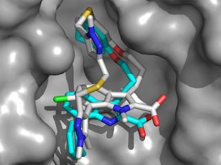
Recognizing the potential synergies of linking these together, the researchers prepared a dimer of a related molecule, but unfortunately the affinity of this much larger molecule was actually worse. However, they wisely isolated and tested a side product, compound 4, and found that this had improved potency. A crystal structure of this molecule bound to Mcl-1 revealed that the pocket had expanded to accommodate the added pyrazole moiety. Since compound 4 adopted a “U-shaped” conformation, the researchers decided to try a macrocyclization strategy to lock this conformation and reduce the entropic penalty of binding. This produced compound 5, and adding a couple more judiciously placed atoms led to AZD5991, with a nearly 300-fold improved affinity. The molecule binds rapidly to Mcl-1 and has a relatively long residence time of about 30 minutes. A crystal structure reveals a close overlay with the initial compound 1 (in cyan).

In addition to picomolar affinity, AZD5991 showed excellent activity in a variety of cancer cell lines dependent on Mcl-1. The compound was tested in mouse and rat xenograft models of multiple myeloma and acute myeloid leukemia and showed complete tumor regression after a single dose. This is all the more remarkable given that AZD5991 is about 25-fold less potent against the mouse version of Mcl-1 than the human version. The molecule was also effective in cell lines resistant to venetoclax, and combining the two molecules caused rapid apoptosis in resistant cell lines. AZD5991 is currently being tested in a phase 1 clinical trial.


This paper holds several lessons. First, the researchers did extensive mechanistic work (beyond the scope of this post to describe) to demonstrate on-target activity. Second, although the initial dimerization strategy was unsuccessful, the researchers turned lemons into lemonade by pursuing a byproduct; we’ve written previously about how even synthetic intermediates are worth testing. Third, the macrocyclization and subsequent optimization is a lovely example of structure-based design and medicinal chemistry. And finally, the fact that the researchers started with a fragment-derived molecule reported by a different group is a testimony to the community nature of science. Last week we highlighted the Open Source Antibiotics initiative, which is actively encouraging others to participate in advancing their early discoveries. Good ideas can come from anywhere, and it takes a lot of them to make a drug.
03 May 06:42

Controlling cellular distribution of drugs with permeability modifying moieties

Med. Chem. Commun., 2019, 10,974-984
DOI: 10.1039/C8MD00412A, Research Article
Paul L. Richardson, Violeta L. Marin, Stormy L. Koeniger, Aleksandra Baranczak, Julie L. Wilsbacher, Peter J. Kovar, Patricia E. Bacon-Trusk, Min Cheng, Todd A. Hopkins, Sandra T. Haman, Anil Vasudevan
Anionic moieties can be used to control the cell-permeability of drugs and used to select the appropriate target identification method for phenotypic screening hits.
The content of this RSS Feed (c) The Royal Society of Chemistry
30 Apr 11:50

Help develop new antibiotics from fragments!

by Dan Erlanson
The state of antibiotic drug discovery is – to put it mildly – dangerously poor. Not only do you have all the challenges inherent to drug discovery, you’re dealing with organisms that can mutate more rapidly than even the craftiest cancer cells. And then there’s the commercial challenge: earlier this month the biotech company Achaogen filed for Chapter 11 bankruptcy, less than a year after winning approval for a new antibiotic.


As Douglas Adams’s Golgafrinchamlearned, complacency about microbial threats is suicidal. But what can any one of us do? Chris Swain, whom we’ve previously highlighted on Practical Fragments, is involved with a consortium of researchers called Open Source Antibiotics. Their mission: “to discover and develop new, inexpensive medicines for bacterial infections.” And they are asking for our help. More on that below.


The researchers initially chose to focus on two essential enzymes necessary for cell wall biosynthesis, MurD and MurE, both of which are highly conserved across bacteria and absent in humans. They conducted a crystallographic fragment screen of both enzymes at XChem, soaking 768 fragments individually at 500 mM concentration. As we’ve writtenpreviously, you’ll almost always get hits if you screen crystallographically at a high enough concentration.


For MurD, four hits were found, all of which bind in the same pocket (in separate structures). Interestingly, this pocket is not the active site, but adjacent to it. The binding modes of the fragments are described in detail here, and the researchers suggest that growing the fragments could lead to competitive inhibitors. The fragments also bind near a loop that has been proposed as a target for allosteric inhibitors, so growing towards this region of the protein would also be an interesting strategy.


MurE was even more productive, with fragments bound at 12 separate sites. (Though impressive, that falls short of the record.) Some of these sites are likely artifacts of crystal packing, or so remote from the active site of the enzyme that they are unlikely to have any functional effects. However, some fragments bind more closely to the active site, and would be good candidates for fragment growing.


If this were a typical publication one might say "cool," and hope that someone picks up on the work sometime in the future. But this, dear reader, is different.


The researchers are actively seeking suggestions for how to advance the hits. Perhaps you want to try running some of these fragments through the Fragment Network? Or do you have a platform, such as “growing via merging,” AutoCouple, or this one, that suggests (and perhaps even synthesizes) new molecules? Perhaps you want to use some of the fragments to work out new chemistry? The consortium has a budget to purchase commercial compounds, and will also accept custom-made molecules. In addition to crystallography, they have enzymatic assays, and are building additional downstream capabilities.


The Centers for Disease Control identifies antibiotic resistance as one of the most serious worldwide health threats. Some have called for a global consortium—modeled after the International Panel for Climate Change—to tackle the problem. But in the meantime, you can play a role yourself. If you would like to participate, you can do so here. The bugs are not waiting for us – and they are already ahead.
29 Apr 20:23

Pharmacological convergence reveals a lipid pathway that regulates C. elegans lifespan

by Alice L. Chen

Pharmacological convergence reveals a lipid pathway that regulates C. elegans lifespan

Pharmacological convergence reveals a lipid pathway that regulates <i>C. elegans</i> lifespan, Published online: 25 March 2019; doi:10.1038/s41589-019-0243-4

Phenotypic screening identifies a small-molecule inhibitor of the C. elegans serine hydrolase FAAH-4 that promotes longevity and identifies the enzyme as the functional homolog of mammalian monoacylglycerol lipase (MAGL).
11 Apr 16:16

Helpful halogens in fragment libraries

by Dan Erlanson
A couple weeks ago we highlighteda small fragment collection (MiniFrags) designed for crystallographic screening. We continue the theme this week with two more papers on the topic, with an emphasis on halogens.


The first, published in J. Med. Chem. by Martin Noble, Michael Waring, and collaborators at Newcastle University, describes a library of “FragLites.” These small (< 14 non-hydrogen atom) fragments are designed to explore “pharmacophore doublets,” such as a hydrogen bond acceptor (HBA) next to a hydrogen bond donor (HBD). For example, the universal fragment 5-bromopyrazole contains an HBA separated by one bond from an HBD. The researchers constructed a set of compounds with either two HBAs or an HBA and an HBD separated by 1 to 5 bonds. Importantly, all compounds also contained either a bromine or iodine atom, the idea being that anomalous dispersion could be used to help identify the fragments using crystallography. A total of 31 FragLites are described, with between 1 to 9 examples for each type of connectivity.


As a test case, these were screened against the kinase CDK2, which has previously been screened crystallographically. FragLites were soaked into crystals at 50 mM, and 9 of the FragLites were found to bind in a total of 6 sites, 4 of which had not been previously observed. The anomalous signal provided by the halogens was important: when the researchers used only normal scattering they identified just 10 of the 16 binding events even when using the powerful PanDDA background correction method. The anomalous signal also helped clarify the binding modes.


The ATP-binding site is where 7 of the 9 FragLites bound, with all but one of them making hydrogen bonding interactions to the hinge region. While not surprising, this does demonstrate that the FragLites can be used experimentally to identify the best binding site. Interestingly, (2-methoxy-4-bromophenyl)acetic acid bound in the active site as well as three other secondary sites; one of these sites hosted three copies of the ligand! It will be interesting to see whether this fragment is generally promiscuous in other proteins too.


As the researchers note, the composition of the FragLite library can be optimized. For example, both of the HBA-HBD fragments with 1-bond separation were identified as hits, while only 3 of the 9 HBA-HBD fragments with 2-bond separation were. Is this due to the choice of fragments, the target tested, or both? The approach is conceptually similar to the Astex minimal pharmacophore concept, so it will be useful to include other types of pharmacophores too (a single HBA or HBD, for example).


A related paper was published in Front. Chem. by Frank Boeckler and colleagues at Eberhard Karls Universität Tübingen. Long-time readers will recallhis earlier halogen-containing library designed for identifying halogen bonds: favorable interactions between halogens and Lewis bases such as carbonyl oxygen atoms. Perhaps because they have relatively stringent geometric requirements (2.75 – 3.5 Å, and a bond angle of 155-180°), halogen bonds are often ignored; the FragLite paper doesn’t even mention them.


The new Boeckler paper describes the construction of a library of 198 halogen-containing fragments, all of which are commercially available and relatively inexpensive. Most of these are rule-of-three compliant, though quite a few also contain more than three hydrogen bond acceptors. Also, given that each fragment contains a halogen, the molecular weights are skewed upward. Solubility was experimentally determined for about half of the fragments, but the highest concentration tested was only 5 mM, and even here several were not fully soluble.


Although no screening data are provided, the researchers note that their “library is available for other working groups.” In the spirit of international cooperation, I suggest a collaboration with the FragLite group!
04 Apr 06:23

A Close Look at Fragments

by Derek Lowe

Here’s a look from the D. E. Shaw research team at fragment binding, and even if you don’t do fragment-based drug discovery, it’s worth a read. That’s because the mechanisms by which fragments bind to proteins are most likely the fundamental ones by which larger molecules bind as well; this is the reductionist look at small molecule-protein interactions. So what kinds of interactions are they?

The group identified 489 fragment-bound protein structures in the PDB with good resolution, with manual inspection to remove the glycerols, etc., from the set. That process also cleared out covalently bound compounds, structures with more than one fragment interacting in the same protein binding pocket, and so on, and left 462 unique fragments and 21 that are bound to more than one protein. That’s a large enough set to draw some conclusions, but it should be noted that the proteins themselves (126 unique ones, with 168 unique binding sites) have hydrolases and transferases over-represented. That doesn’t mean that the interactions detected are any less valid, but you wouldn’t want to necessarily depend on a raw count of them (the authors normalized the data to deal with this problem). Most of the compound set has between 10 and 16 heavy atoms, most are uncharged, and most have cLogP values less than 2. Of the number of ring assemblies in the structures, phenyl, pyridine, pyrazole, thiophene, and indole account for half the numerical total, but of the 178 unique ring assemblies, 60% of them occur only once in the set.

What about binding? The large majority bury more than 80% of their solvent-accessible surface area when bound, which is what you’d expect from such small molecules that are able to display good potency for their size. The least-buried compound in the whole set is still at 50%. And if you consider polar surface area, a bit over 50% of the compounds bury over 90% of their polarity when bound, which makes sense, too – you’re not going to get noticeable binding at these molecular sizes just by random hydrophobic interactions alone. And indeed, 92% of the structures have at least one hydrogen bond to the protein, to a structural water molecule, or to a metal atom (such as a zinc in an active site, which is what you see with, in the classic example, sulfonamides bound to carbonic anhydrase). The record-holder is this structure, with 7 such interactions (!), followed by this one with six hydrogen bonds alone.

Considering the amount of time some of us has spent trying to get one measly hydrogen bond into our complexes, that’s pretty impressive. As experienced medicinal chemists know, those things are wonderful for picking up enthalpic currency in binding, but they’re extremely finicky (profoundly directional, for one thing). And as been often noted, an unsuccessful attempt at adding a hydrogen bond generally leaves you worse off than before, with a polar group that has had to shed its solvent interactions for no particular return. And that’s one of the principles behind a fragment-based approach – you try to start out with a core that already is displaying these features. The paper goes into detail on the varieties of nitrogen and oxygen atoms that participate in these hydrogen bonds from the fragment structures, and the sorts of groups on the protein on the other side of the transaction.

The biggest category of interaction outside of the hydrogen bonds are general arene-ring interactions, at 42% of the examples. That’s often called “pi-stacking” by Cro-Magnons like me, but it also encompasses edge-to-pi, arene-to-cation, and other categories. And the third most common category, found in 12% of the examples, is actually C-H hydrogen bonds, which don’t get nearly as much attention.

Overall, the paper recommends that if you want to generate new fragment libraries, that you stick with about a quarter of the heavy atoms being polar ones capable of participating in hydrogen bonding – in practice, that pretty much means nitrogen and oxygen atoms. Amides and alcohols are particularly useful, since they can both accept and donate in this context. You should keep things simple and not try to decorate the fragments with too many pharmacophores, because the simpler compounds have more geometric freedom to find productive interactions without running into additional clashes. The paper proposes that there must be a number of lesser-used heterocycles that could both participate in hydrogen bonds and in arene pi interactions, and they also suggest that seven-membered rings are under-represented, especially considering their unique conformational properties. But if you’re interested in having a rough first-pass set of fragments, though, to assess druggability and the like, you could do far worse than picking as many of the 462 compounds in this paper’s set as you can get. They surely have some structural biases from the earlier days of commercial fragment libraries, but they also have a proven record of binding to proteins.

The tricky part of putting new or unusual fragment structures into your library is that you have to be able to functionalize them later on, and ideally in several possible directions. I ran into that myself a few years back – a rather underexplored ring system came up (cinnolines) that really bound quite well to the protein target. But the number of known methods to make a variety of functionalized cinnolines is limited, so you have to decide if you want to embark on a discover-new-chemistry project in order to embark on the develop-a-fragment project in turn.

Interestingly, considering that this paper is coming from one of the most high-powered modeling groups in the business, the point is made that the number of water interactions in the studied set presents a problem. Current modeling software does not handle these things as well as it does some other categories of interaction, so virtual fragment screening efforts are going to have significant blind spots. This goes, of course, both for more static docking approaches and for molecular dynamics. Another computational issue is the ability to find and picture nonbonded interactions – as it stands, there seems to be too much handwork involved with querying the PDB for such things.

Overall, the paper is both a hymn to fragment-based drug discovery and (less directly) one to crystallography. It’s X-ray data that underpin the whole thing (and underpins the vast majority of fragment-based drug work in general). That really is the ground truth of this approach, for all the known limitations of crystallographic data – not modeling, not simulations. And the more of it we can get, the more we’ll understand what we’re doing.

26 Mar 22:33

Tiny fragments at high concentrations give massive hit rates

by Dan Erlanson
Screening fragments crystallographically is becoming more common, especially as the process becomes increasingly automated. Not only does crystallography reveal detailed molecular contacts, it is unmatched in sensitivity. At the FBLD 2018 meeting last year we highlighted work out of Astex taking this approach to extremes, screening very small fragments at very high concentrations. Harren Jhoti and colleagues have now published details (open access) in Drug Discovery Today.


The researchers assembled a library of 81 diminutive fragments, or “MiniFrags”, each with just 5 to 7 non-hydrogen atoms. Indeed, the fragments adhere more closely to the “rule of 1” than the “rule of 3.” Because the fragments are so small, they are likely to have especially low affinities: a 5 atom fragment with an impressive ligand efficiency of 0.5 kcal mol-1 per heavy atom would have a risibly weak dissociation constant of 14 mM. In order to detect such weak binders, the researchers screen at 1 M fragment concentrations, almost twice the molarity of sugar in soda! Achieving these concentrations is done by dissolving fragments directly in the crystallographic soaking solution and adjusting the pH when necessary. Although this might mean preparing custom fragment stocks for each protein, it avoids organic solvents such as DMSO, which can both damage crystals and compete for ligand binding sites.


As proof of concept, the researchers chose five internal targets they had previously screened crystallographically under more conventional conditions (50-100 mM of larger fragments). All targets diffracted to high resolution, at least 2 Å, and represented a range of protein classes from kinases to protein-protein interactions. The hit rates were enormous, from just under 40% to 60%, compared to an average of 12% using standard conditions.


Astex has previously described how crystallography often identifies secondary binding sites away from the active site, and this turned out to be the case with MiniFrags: an average of 10 ligand binding sites per protein. In some cases protein conformational changes occurred, which is surprising given the small size and (presumably) weak affinities of the MiniFrags.


All this is fascinating from a molecular recognition standpoint, but the question is whether it is useful for drug discovery. The researchers go into some detail around the kinase ERK2, which we previously wrote about here. MiniFrags identified 11 ligand-binding sites, several of which consist of subsites within the active site. Some of the MiniFrags show features previously seen in larger molecules, such as an aromatic ring or a positively charged group, but the MiniFrags also identified new pockets where ligands had not previously been observed. The researchers argue that these “warm spots” could be targeted during lead optimization.


One laudable feature of the paper is that the chemical structures of all library members are provided in the supplementary material. Although it would be easy to recreate by purchasing compounds individually, hopefully one or more library vendors will start selling the set. If MiniFrag screening is standardized across multiple labs, the resulting experimental data could provide useful inputs for further improving computational approaches, as well as providing more information for lead discovery.
13 Feb 15:34

[ASAP] Fragment-Based Covalent Ligand Screening Enables Rapid Discovery of Inhibitors for the RBR E3 Ubiquitin Ligase HOIP

by Henrik Johansson, Yi-Chun Isabella Tsai, Ken Fantom, Chun-Wa Chung, Sandra Kümper, Luigi Martino, Daniel A. Thomas, H. Christian Eberl, Marcel Muelbaier, David House, Katrin Rittinger

TOC Graphic

Journal of the American Chemical Society
DOI: 10.1021/jacs.8b13193
28 Jan 23:36

Science And Society—What Do They Owe Each Other?

by Gautam R. Desiraju
Angewandte Chemie International Edition Science And Society—What Do They Owe Each Other?

Scientists owe it to society to provide guidance, leadership, and above all, moral stature, so that they become role models… Both knowledge and wealth are acquired only through the pursuit of truth. This is smoothly achieved when then there is a seamless exchange of unspoken thought between science and society. …” Read more in the Guest Editorial by G. R. Desiraju.


28 Jan 23:35

Selective and reversible modification of kinase cysteines with chlorofluoroacetamides

by Naoya Shindo

Selective and reversible modification of kinase cysteines with chlorofluoroacetamides

Selective and reversible modification of kinase cysteines with chlorofluoroacetamides, Published online: 14 January 2019; doi:10.1038/s41589-018-0204-3

Discovery and exploitation of inherent reaction features of chlorofluoroacetamide (CFA) as a warhead such as low off-target activity and reversible reactivity with cysteine enable specific covalent inhibition of targeted kinases.
10 Dec 19:45

Recent Applications of Diazirines in Chemical Proteomics

by Jean-Philip George Lumb, Matthew W. Halloran
Chemistry – A European Journal Recent Applications of Diazirines in Chemical Proteomics

The elucidation of substrate–protein interactions is an important component of the drug development process. Photoaffinity labeling (PAL) is a versatile technique that can provide insight into ligand–target interactions. Among the commonly employed photoreactive groups, diazirines have emerged as the gold standard. In this Minireview, recent developments in the field of diazirine‐based PAL are discussed, with emphasis on their applications in proteomic studies.


Abstract

The elucidation of substrate–protein interactions is an important component of the drug development process. Due to the complexity of native cellular environments, elucidating these fundamental biochemical interactions remains challenging. Photoaffinity labeling (PAL) is a versatile technique that can provide insight into ligand‐target interactions. By judicious modification of substrates with a photoreactive group, PAL creates a covalent crosslink between a substrate and its biological target following UV‐irradiation. Among the commonly employed photoreactive groups, diazirines have emerged as the gold standard. In this Minireview, recent developments in the field of diazirine‐based photoaffinity labeling will be discussed, with emphasis being placed on their applications in chemical proteomic studies.

10 Dec 19:25

[ASAP] Photogeneration of Quinone Methides as Latent Electrophiles for Lysine Targeting

by Raúl Pérez-Ruiz, Oscar Molins-Molina, Emilio Lence, Concepción González-Bello, Miguel A. Miranda, M. Consuelo Jiménez

TOC Graphic

The Journal of Organic Chemistry
DOI: 10.1021/acs.joc.8b01559
08 Nov 15:53

[ASAP] Binding Kinetics Survey of the Drugged Kinome

by Victoria Georgi, Felix Schiele, Benedict-Tilman Berger, Andreas Steffen, Paula A. Marin Zapata, Hans Briem, Stephan Menz, Cornelia Preusse, James D. Vasta, Matthew B. Robers, Michael Brands, Stefan Knapp, Amaury Fernández-Montalván

TOC Graphic

Journal of the American Chemical Society
DOI: 10.1021/jacs.8b08048