Shared posts

13 Jan 05:23

Assembly of black phosphorus quantum dots-doped MOF and silver nanoclusters as a versatile enzyme-catalyzed biosensor for solution, flexible substrate and latent fingerprint visual detection of baicalin

Publication date: 15 March 2020

Source: Biosensors and Bioelectronics, Volume 152

Author(s): Xiaowen Jiang, Hui Jin, Yujiao Sun, Zejun Sun, Rijun Gui

Abstract

In this work, a versatile enzyme-catalyzed biosensor was developed by using the assembled nanohybrids of black phosphorus quantum dots (BPQDs)-doped metal-organic frameworks (MOF) and silver nanoclusters (AgNCs). The nanohybrids of AgNCs/BPQDs/MOF exhibit dual-emissive fluorescence (FL) centers at 630 nm (red) and 535 nm (blue) under excitation at 440 nm. Baicalin enhances the activity of catalase and catalytic decomposition of H2O2. With increase of baicalin contents in the mixture containing nanohybrids, catalase and H2O2, the catalase-caused decomposition of H2O2 was accelerated and the excessive H2O2 was consumed. Baicalin can restrain the oxidation capability of H2O2. The red-FL (response signal) of AgNCs adhering to MOF increases, while blue-FL (reference signal) of BPQDs doped into MOF has negligible changes. A new ratiometric FL biosensor was designed based on nanohybrids and enzyme-catalyzed reaction. This biosensor enables the detection of baicalin in the range of 0.01–500 μg mL−1, with a limit of detection of 3 ng mL−1. This biosensor has high sensitivity, selectivity and stability for baicalin detection in practical samples. Especially, it performed the solution, flexible substrate and latent fingerprint visual detection of baicalin through direct observation of FL color shades with naked eyes. This work explored a facile and efficient semi-quantitative method for versatile FL visual detection, which can promote the development of advanced chemo/bio-sensors and analysis methods.

Graphical abstract

Image 1

16 Jul 02:38

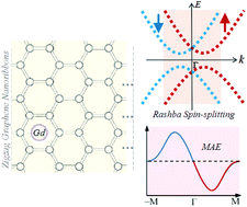
Unconventional magnetic anisotropy in one-dimensional Rashba system realized by adsorbing Gd atom on zigzag graphene nanoribbons

Nanoscale, 2017, 9,11657-11666
DOI: 10.1039/C7NR03164E, Paper
Zhenzhen Qin, Guangzhao Qin, Bin Shao, Xu Zuo
The Rashba effect, a spin splitting in electronic band structure, can be induced to the graphene nanoribbon by the transverse electronic field due to the asymmetric adsorption of Gd atom, which would impact the magnetic anisotropy distribution in k-space.
The content of this RSS Feed (c) The Royal Society of Chemistry
02 Aug 06:20

Rapid and Energy-Saving Microwave-Assisted Solid-State Synthesis of Pr3+-, Eu3+-, or Tb3+-Doped Lu2O3 Persistent Luminescence Materials

by Cássio C. S. Pedroso, José M. Carvalho, Lucas C. V. Rodrigues, Jorma Hölsä and Hermi F. Brito

TOC Graphic

ACS Applied Materials & Interfaces
DOI: 10.1021/acsami.6b04683
12 Jan 13:35

Tunable nanogap devices for ultra-sensitive electrochemical impedance biosensing

Publication date: 28 January 2016
Source:Analytica Chimica Acta, Volume 905
Author(s): Yong Lu, Zheng Guo, Jing-Jing Song, Qin-An Huang, Si-Wei Zhu, Xing-Jiu Huang, Yan Wei
A wealth of research has been available discussing nanogap devices for detecting very small quantities of biomolecules by observing their electrical behavior generally performed in dry conditions. We report that a gold nanogapped electrode with tunable gap length for ultra-sensitive detection of streptavidin based on electrochemical impedance technique. The gold nanogap is fabricated using simple monolayer film deposition and in-situ growth of gold nanoparticles in a traditional interdigitated array (IDA) microelectrode. The electrochemical impedance biosensor with a 25-nm nanogap is found to be ultra-sensitive to the specific binding of streptavidin to biotin. The binding of the streptavidin hinder the electron transfer between two electrodes, resulting in a large increase in electron-transfer resistance (R et) for operating the impedance. A linear relation between the relative R et and the logarithmic value of streptavidin concentration is observed in the concentration range from 1 pM (picomolar) to 100 nM (nanomolar). The lowest detectable concentration actually measured reaches 1 pM. We believe that such an electrochemical impedance nanogap biosensor provides a useful approach towards biomolecular detection that could be extended to a number of other systems.

Graphical abstract

image